


Computed tomography (CT or CAT scan) ranks as one of the top five medical developments in the last 50 years, according to most medical surveys. CT has proven so valuable as a medical diagnostic tool that the 1979 Nobel Prize in Medicine was awarded to the inventors.
Certain medical conditions require a deeper look, usually at the tissues, blood vessels, and bones inside your body. X-rays and ultrasounds can provide some information, but when a more detailed view is required, a computed tomography (CT) scan is usually the next step.
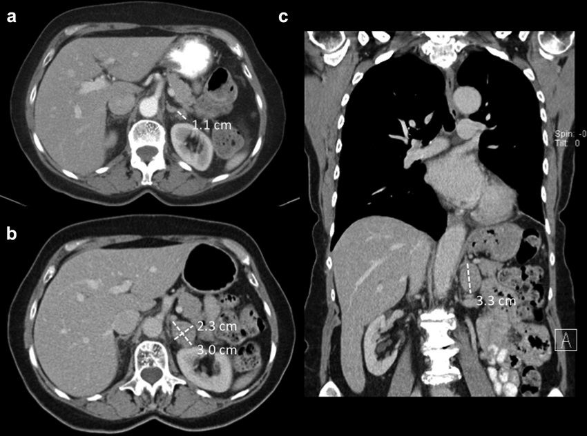
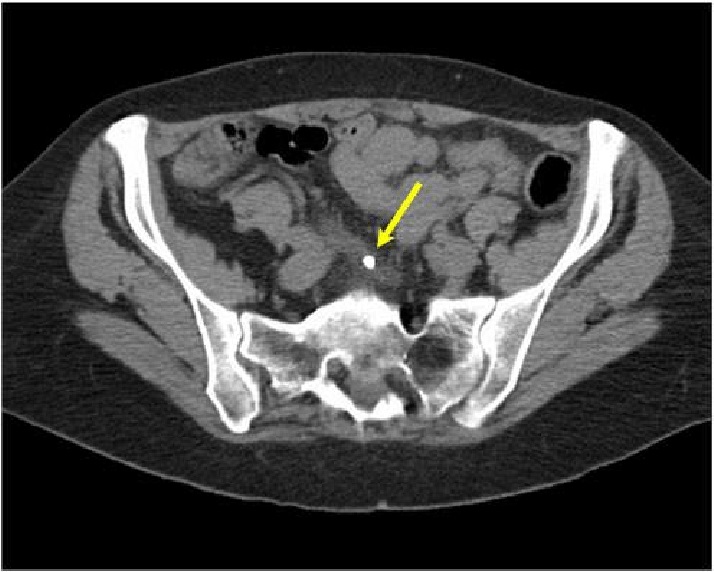
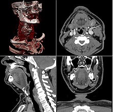
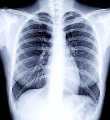
A CT scan uses computers and rotating X-ray machines to create cross-sectional images of the body. These images provide more detailed information than typical X-ray images. They can show the soft tissues, blood vessels, and bones in various parts of the body.
A CT scan has many uses, but it’s particularly well-suited for diagnosing diseases and evaluating injuries. The imaging technique can help your doctor:
CT scans don’t require much preparation. If needed, you can do a CT scan with or without contrast very quickly.
If you are scheduled for a CT scan with contrast dye, it may help to refrain from eating solid foods for up to 4 hours before your test.
If your doctor is using oral contrast for your CT scan, you’ll probably be given the contrast before the day of your scan and instructed on how to prepare and drink it. Generally, you will want to start drinking the solution within an hour or two of your scan, drinking a portion of the solution every 15 minutes.
Otherwise, the only preparations you need to take before a CT scan are to remove metallic objects and medication devices from your body. This includes:
When you arrive for your CT scan, you’ll be asked to change into a hospital gown. The technician doing your scan may insert an IV catheter in your arm or leg and ask whether you have removed any metal devices or medication patches prior to your arrival.
When it’s time to begin the scan, you’ll be positioned on a long narrow table.The technician will leave the room before operating the scanner and may give you instructions over an intercom.
As the table moves in and out of the scanner, the machine will rotate around you making a loud noise. You may be asked to hold your breath or maintain certain positions. Otherwise, you should hold as still as possible to prevent the scanner from capturing blurry images.
The entire process should take between 20 minutes and 1 hour.
The CT scan itself is a painless procedure. Some people feel uncomfortable on the hard table or have difficulty remaining still.
You may feel a slight burning when the contrast dye enters your vein. Some people experience a metal taste in their mouths and a warm sensation throughout their bodies. These reactions are normal and generally last less than a minute.
A radiologist, a doctor trained to supervise and interpret radiology exams, will analyze the images. The radiologist will send a signed report to your primary care or referring physician, who will share the results with you.
Benefits of CT include more effective medical management by:
The risks are based on the type of imaging as well as how the imaging is performed.
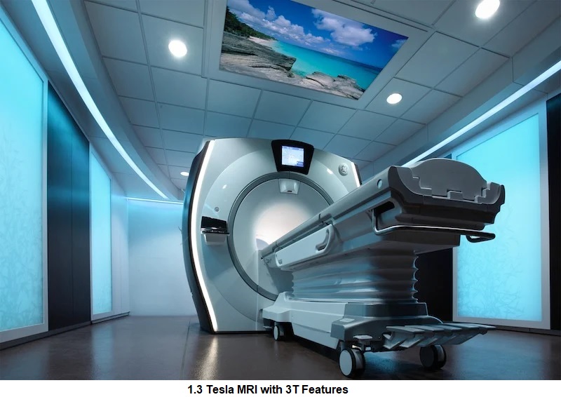
MRI stands for Magnetic Resonance Imaging. MRI uses the principle of magnetic resonance. The hydrogen protons inside our body are magnetizable when placed in a large magnet, like the one in the MRI machine. When the body part goes into the machine, these hydrogen protons get magnetized. Using this property, images are produced of different parts of the body.
Magnetic resonance imaging (MRI) is a noninvasive test doctors use to diagnose medical conditions.
MRI uses a powerful magnetic field, radiofrequency pulses, and a computer to produce detailed pictures of internal body structures. MRI does not use radiation (x-rays).
Detailed MR images allow doctors to examine the body and detect disease.
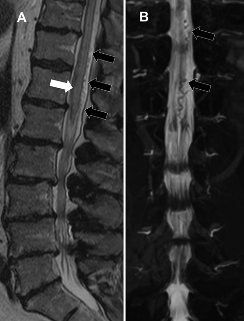
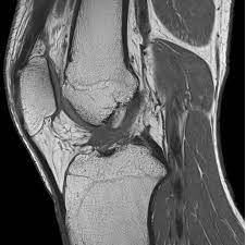
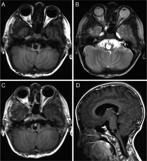
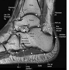
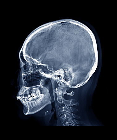
MRI allows us to exquisitely see different parts of the body. In the brain, it allows us to differentiate between different areas. In the spine, MRI is the only modality that allows us to see the spinal cord. In joints, MRI allows us to view the internal parts of the joints, including ligaments, tendons and menisci. It also allows evaluation of various cardiac structures, as well as functional imaging of various parts of the brain. MRI does all this by sectioning the body in different planes. Just as it is necessary to slice a loaf of bread to know the quality of the slices, so also, MRI allows us to section the body to view its inner parts. Thus, the skull X-ray only shows us the outer part, whereas MRI shows us the inner parts of the brain.
You will need to change into a hospital gown. This is to prevent artifacts appearing on the final images and to comply with safety regulations related to the strong magnetic field.
Guidelines about eating and drinking before an MRI vary between specific exams and facilities. Take food and medications as usual unless your doctor tells you otherwise.
Some MRI exams use an injection of contrast material. The doctor may ask if you have asthma or allergies to contrast material, drugs, food, or the environment.
Tell the technologist or radiologist if you have any serious health problems or recent surgeries. Some conditions, such as severe kidney disease.You may need a blood test to confirm your kidneys are functioning normally.
Women should always tell their doctor and technologist if they are pregnant. MRI has been used since the 1980s with no reports of any ill effects on pregnant women or their unborn babies. However, the baby will be in a strong magnetic field. Therefore, pregnant women should not have an MRI in the first trimester unless the benefit of the exam clearly outweighs any potential risks.
Leave all jewelry and other accessories at home or remove them prior to the MRI scan. Metal and electronic items are not allowed in the exam room. They can interfere with the magnetic field of the MRI unit, cause burns, or become harmful projectiles. These items include:
In most cases, an MRI exam is safe for patients with metal implants, except for a few types. People with the following implants may not be scanned and should not enter the MRI scanning area without first being evaluated for safety:
Infants and young children often require anesthesia to complete an MRI exam without moving. This depends on the child's age, intellectual development, and the type of exam. A specialist in pediatric anesthesia should be available during the exam for your child's safety. You will be told how to prepare your child.
Most MRI exams are painless. However, some patients find it uncomfortable to remain still. Others may feel closed-in (claustrophobic) while in the MRI scanner. The scanner can be noisy.
It is important that you remain perfectly still while the images are being taken. This is typically only a few seconds to a few minutes at a time. You will know when images are being recorded because you will hear and feel loud tapping or thumping sounds.
You will be provided with earplugs or headphones to reduce the noise made by the scanner. You may be able to relax between imaging sequences. However, you will need to keep the same position as much as possible without moving.
You will usually be alone in the exam room. However, the technologist will be able to see, hear, and speak with you at all times using a two-way intercom.
In some cases, IV injection of contrast material may be given before the images are obtained. The IV needle may cause you some discomfort.
There is also a very small chance of skin irritation at the site of the IV tube insertion. Some patients may have a temporary metallic taste in their mouth after the contrast injection.
You may resume your usual activities and normal diet immediately after the exam. On very rare occasions, a few patients experience side effects from the contrast material.These may include nausea, headache, and pain at the site of injection.
A radiologist, a doctor trained to supervise and interpret radiology exams, will analyze the images. The radiologist will send a signed report to your primary care or referring physician, who will share the results with you.
High-quality images depend on your ability to remain perfectly still and follow breath-holding instructions while the images are being recorded. If you are anxious, confused or in severe pain, you may find it difficult to lie still during imaging.
A person who is very large may not fit into certain types of MRI machines. There are weight limits on the scanners.
Implants and other metallic objects can make it difficult to obtain clear images. Patient movement can have the same effect.
A very irregular heartbeat may affect the quality of images. This is because some techniques time the imaging based on the electrical activity of the heart.
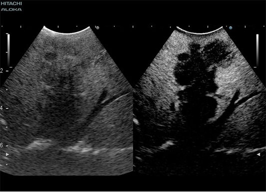
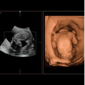
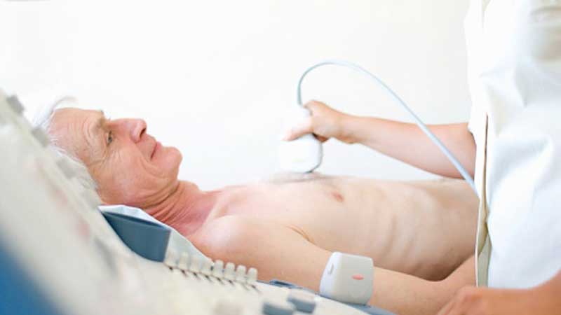
Ultrasound is a noninvasive imaging test that shows structures inside your body using high-intensity sound waves. Healthcare providers use ultrasound exams for several purposes, including during pregnancy, for diagnosing conditions and for image guidance during certain procedures.
It helps diagnose the causes of pain, swelling and infection in the body's internal organs and to examine an unborn child (fetus) in pregnant women. It also helps guide biopsies, diagnose heart conditions, and assess damage after a heart attack. Ultrasound is safe, noninvasive, and does not use radiation.
This procedure requires little to no special preparation. Your doctor will tell you how to prepare, including whether you should not eat or drink beforehand.
Ultrasound exams can help diagnose a variety of conditions and assess organ damage following illness. Doctors use ultrasound to evaluate:
Ultrasound is a useful way of examining many of the body's internal organs, including but not limited to the:
Ultrasound is also used to:
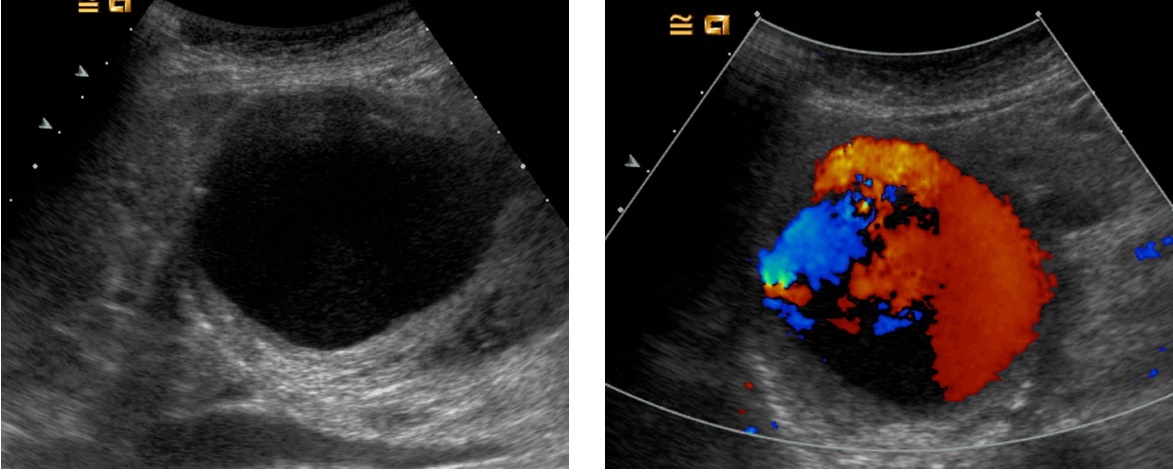
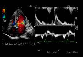
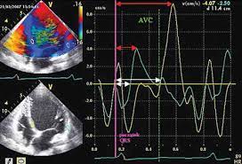
Doppler ultrasound helps the doctor to see and evaluate:
With knowledge about the speed and volume of blood flow gained from a Doppler ultrasound image, the doctor can often determine whether a patient is a good candidate for a procedure like angioplasty.
Wear comfortable, loose-fitting clothing. You may need to remove all clothing and jewelry in the area to be examined. You may need to change into a gown for the procedure.
Most ultrasound exams require no preparation. However, there are a few exceptions:
A radiologist, a doctor trained to supervise and interpret radiology exams, will analyze the images. The radiologist will send a signed report to your primary care or referring physician, who will share the results with you.
Ultrasound waves are disrupted by air or gas. Therefore, ultrasound is not an ideal imaging technique for the air-filled bowel or organs obscured by the bowel. Ultrasound is not as useful for imaging air-filled lungs, but it may be used to detect fluid around or within the lungs. Similarly, ultrasound cannot penetrate bone, but may be used for imaging bone fractures or for infection surrounding a bone.
Large patients are more difficult to image by ultrasound because greater amounts of tissue weaken the sound waves as they pass deeper into the body and need to return to the transducer for analysis.
Ultrasound has difficulty penetrating bone and, therefore, can only see the outer surface of bony structures and not what lies within (except in infants who have more cartilage in their skeletons than older children or adults). Doctors typically use other imaging modalities such as MRI to visualize the internal structure of bones or certain joints.
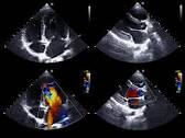
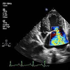
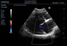
An echocardiogram (echo) is a test that uses high frequency sound waves (ultrasound) to make pictures of your heart. The test is also called echocardiography or diagnostic cardiac ultrasound.
An echo uses sound waves to create pictures of your heart’s chambers, valves, walls and the blood vessels (aorta, arteries, veins) attached to your heart. A probe called a transducer is passed over your chest. The probe produces sound waves that bounce off your heart and “echo” back to the probe. These waves are changed into pictures viewed on a video monitor. An echo can’t harm you.
Your doctor may use an echo test to look at your heart’s structure and check how well your heart functions. The test helps your doctor find out:
The test also will help your doctor find out if there are:
You don’t have to do anything special. You can eat and drink before the test like you usually would.
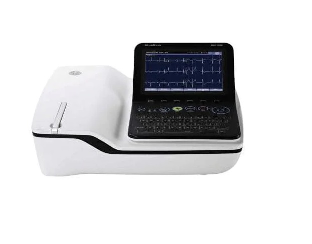
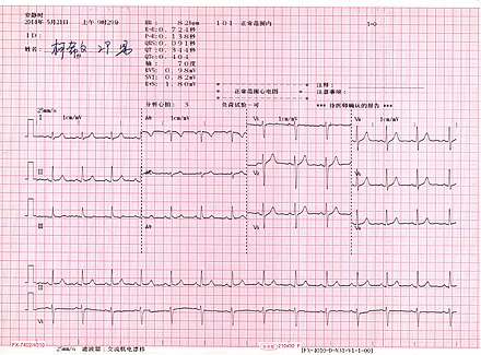
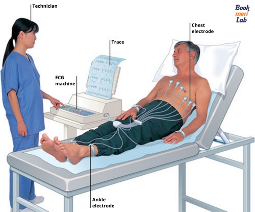
An ECG or EKG is an imaging procedure done by a technician to access the heart wave activity or to monitor the status of the heart’s electrical conduction system. It is the first imaging tests recommended to the patients suffering from chest pain or other heart symptoms.
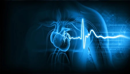
In simpler, an electrocardiogram is a graphical presentation of the electrical activity of the heart muscles. The wavy lines series in an ECG report are the readings of an ECG machine that records the tracings of heartbeats and prints them onto paper. These readings are tools to diagnose heart disorders.
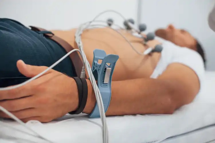
ECGs are the diagnostic tools that help the technician look for underlying heart conditions such as chest pain, an irregular heartbeat, shortness of breath or heavy heartbeats. Most cardiologists use ECGs to assesses the heart rhythm, chamber size, and muscle thickness. The following are some of the common reasons for which for your doctor may request an electrocardiogram (ECG):
An electroencephalogram (EEG) is a test used to evaluate the electrical activity in your brain. Brain cells communicate with each other through electrical impulses. An EEG can be used to help detect potential problems associated with this activity.
An EEG tracks and records brain wave patterns. Small flat metal discs called electrodes are attached to your scalp with wires. The electrodes analyze the electrical impulses in your brain and send signals to a computer that records the results.
An EEG is one of the main diagnostic tests for epilepsy. An EEG can also play a role in diagnosing other brain disorders.
An EEG can find changes in brain activity that might be useful in diagnosing brain disorders, especially epilepsy or another seizure disorder. An EEG might also be helpful for diagnosing or treating:
Ask your doctor if you should stop taking any medications before the test. Wash your hair the night before the EEG. Don’t put any products like sprays or gels on the day of the test. Avoid eating or drinking anything containing caffeine for at least 8 hours before the test.
Electromyography (EMG) is a diagnostic procedure that evaluates the health condition of muscles and the nerve cells that control them. EMG results can help diagnose muscle disorders, nerve disorders, and disorders affecting the connection between nerves and muscles.
Your doctor may order an EMG if you have signs or symptoms that may indicate a nerve or muscle disorder. Such symptoms may include:
EMG results are often necessary to help diagnose or rule out a number of conditions such as:
A nerve conduction velocity (NCV) test is used to assess nerve damage and dysfunction. Also known as a nerve conduction study, the procedure measures how quickly electrical signals move through your peripheral nerves.
An NCV test can be used to diagnose a number of muscular and neuromuscular disorders, including:
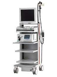
Endoscopy is the insertion of a long, thin tube directly into the body to observe an internal organ or tissue in detail. It can also be used to carry out other tasks, including imaging and minor surgery.
An upper endoscopy, also called an upper gastrointestinal endoscopy, is a procedure used to visually examine your upper digestive system. This is done with the help of a tiny camera on the end of a long, flexible tube. A specialist in diseases of the digestive system (gastroenterologist) uses an endoscopy to diagnose and sometimes treat conditions that affect the upper part of the digestive system.
You may need an upper endoscopy if you have unexplained:
For many types of endoscopy, the individual needs to fast for around 12 hours, though this varies based on the type. A doctor will conduct an examination before the endoscopy. It is important to mention all current medications (including supplements) and any previous procedures.
A colonoscopy is an outpatient procedure that is done to examine the inside of your large intestine (colon and rectum). The examination uses an instrument called a colonoscope (sometimes called a scope). This flexible instrument is very long and includes a camera and the ability to remove tissue (you won't feel the tissue being removed). A colonoscopy is commonly used to evaluate gastrointestinal symptoms, such as bleeding, abdominal pain or changes in bowel habits (how often you poop, how easily you poop, and the color and consistency of your poop).
Your doctor may recommend a colonoscopy to:
- Investigate intestinal signs and symptoms
- Screen for colon cancer
- Look for more polyps
- Treat an issue
Before a colonoscopy, you'll need to clean out (empty) your colon.
There are a few different kinds of bowel preparations for colonoscopy, almost all of them liquid. Your doctor will tell you what kind is best for you based on your medical history and their particular preference.
The time of day or night that you will have to start drinking the solution will depend on when your procedure is scheduled. You will be asked to consume the entire amount of liquid within a specific time period.
Be sure to drink plenty of fluids the day before while you are doing your bowel prep.
You should not drink or eat anything at all for at least four hours before the colonoscopy.
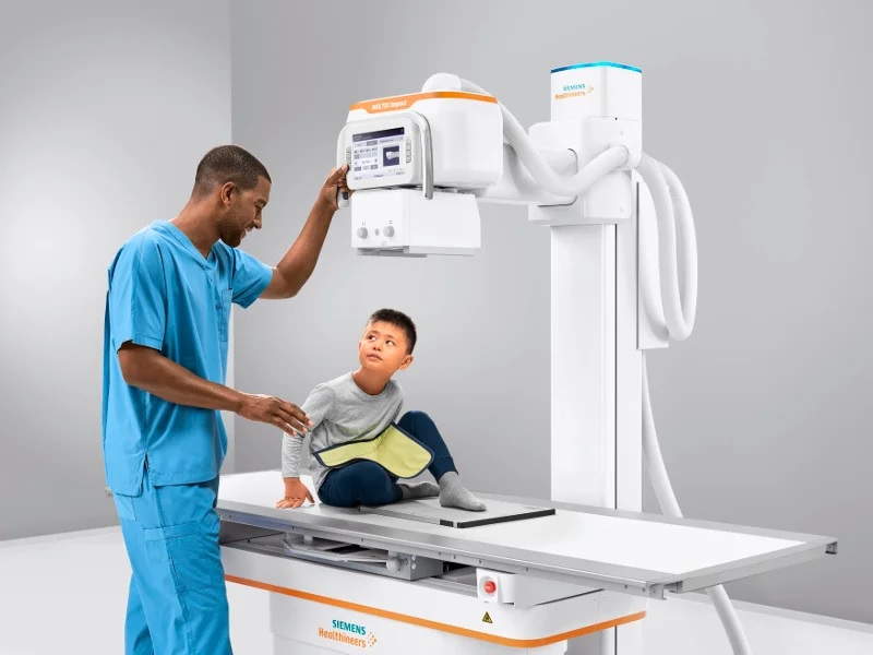
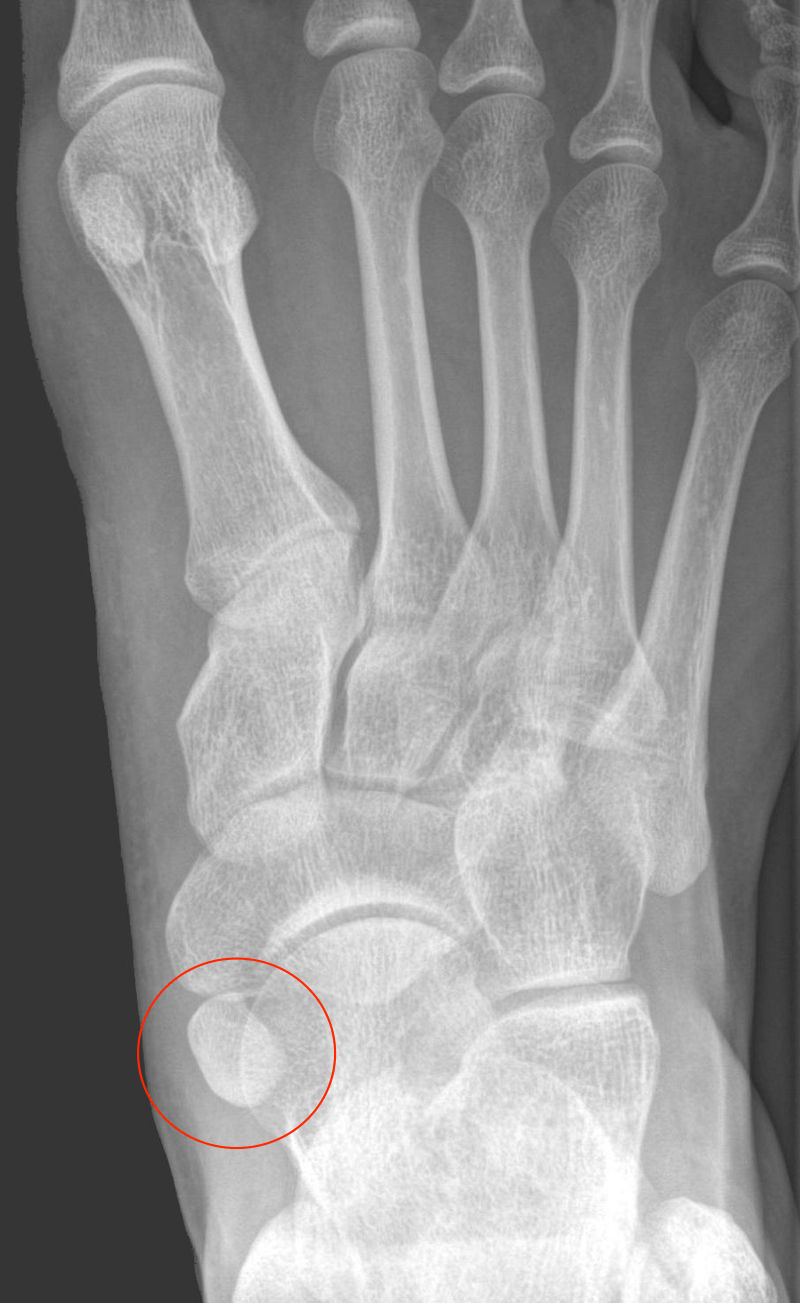
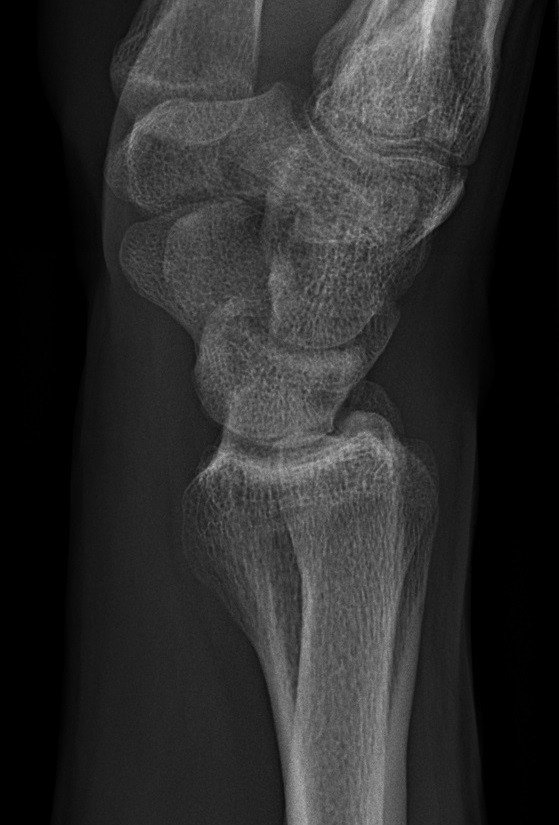
An X-ray is an imaging study that takes pictures of bones and soft tissues. X-rays use safe amounts of radiation to create these pictures. The images help healthcare providers diagnose a wide range of conditions and plan treatments. Usually, providers use X-rays to evaluate broken bones, dislocated joints and other bone injuries.
An X-ray study (also called a radiograph) is a type of medical imaging (radiology) that creates pictures of your bones and soft tissues, such as organs. X-rays use safe amounts of radiation to make these pictures. The images help your provider to diagnose conditions and plan treatments.
Most often, providers use X-rays to look for fractures (broken bones). But X-ray images can help providers diagnose a wide range of injuries, disorders and diseases. X-rays are a safe and effective way for providers to evaluate your health.
Your doctor may order an X-ray to:
Conditions that may call for an X-ray include:
X-rays are standard procedures. In most cases, you won’t need to take special steps to prepare for them. Depending on the area that your doctor and radiologist are examining, you may want to wear loose, comfortable clothing that you can easily move around in. They may also ask you to remove any jewelry or other metallic items from your body before your X-ray is taken.
Always tell your doctor or radiologist if you have metal implants from prior surgeries. These implants can block X-rays from passing through your body and creating a clear image.
In some cases, you may need to take a contrast material or “contrast dye” before your X-ray. This is a substance that will help improve the quality of the images.
If you’re having an X-ray to examine your gastrointestinal tract, your doctor may ask you to fast for a certain amount of time beforehand. You will need to avoid eating anything while you fast. You may also need to avoid or limit drinking certain liquids. In some cases, they may also ask you to take medications to clear out your bowels.
X-rays use small amounts of radiation to create images of your body. The level of radiation exposure is considered safe for most adults, but not for a developing baby. If you’re pregnant or believe you could be pregnant, tell your doctor before you have an X-ray. They may suggest a different imaging method, such as an MRI.
If you ingest a contrast material before your X-ray, it may cause side effects. These include:
Fine needle aspiration cytology or an FNAC is a diagnostic test that is used to check if a lump or a mass on the body is cancerous in nature. As the name suggests, this test is performed using a needle.
FNAC test is performed to determine the nature of superficial lumps- both palpable (can be felt on touch) and impalpable (can’t be felt easily on touch), such as those on breast or neck.
It is specifically recommended when an abnormal lump is felt or detected during other diagnostic and routine medical procedures.
No prior preparations are needed for performing this test.
Fine Needle Aspiration Cytology is the abbreviation for Fine Needle Aspiration Cytology. The FNAC test is a simple, rapid, and low-cost test used to assess a specific condition or body region. It involves aspirating cells from a particular region of the body using a fine gauge needle. The sample is then sent to the lab, where it is examined under a microscope through several processes. This test is quite helpful in diagnosing inflammatory diseases and malignancies of varying types. This technique is commonly used for examination of the breast, kidney, liver, lungs, prostate, pancreas, salivary glands, retroperitoneum, and lymph nodes.
Histopathology is the study of tissues related to disease. Histopathology is the diagnosis and study of diseases of the tissues, and involves examining tissues and/or cells under a microscope. Histopathologists are responsible for making tissue diagnoses and helping clinicians manage a patient’s care.
 Doctors most commonly perform histopathology so that a histopathologist can view the sample for the presence of cancerous cells. Some of the key questions a histopathologist may report on can include:
Haematology is referred to the study, diagnosis and management of blood related diseases such asanemia, infection, haemophilia, blood-clotting disorders, and leukaemia.
Cytology is the microscopic examination of cell samples. These samples can be collected from any area of the body. Cytology is often used to diagnose growths or masses (tumors) found on the surface of the body, but can also be used to assess bodily fluids, internal organs (e.g., liver, lung, lymph nodes, kidney), and abnormal fluids that may accumulate, especially in the chest and abdomen.
There are several ways to collect the cells depending on where the problem is and what type of tissue is involved.
Most commonly, fine needle aspiration or fine needle biopsy is performed to collect cells.
Other techniques may be used, such as: Skin scraping, Impression smear, Cotton-tipped swabs and Lavage etc.
Cytology is less diagnostic than histopathology. Histopathology is the examination of samples of whole tissues and is performed on a solid piece of tissue that has been collected surgically.Histopathology focuses on the architecture of the tissue and provides more information about the tissue than cytology.
Clinical Pathology is the branch of pathology directed to the diagnosis and monitoring of diseases via examination of blood, bodily fluids, secretions, and biopsy specimens. The Clinical Pathology section evaluates these specimens for chemical, morphologic, and immunologic abnormalities. A variety of test procedures and microscopic examinations are performed to confirm a clinical impression, establish or exclude a diagnosis, monitor therapy, and help establish a prognosis.
Microbiology deals with microscopic organisms like viruses, bacteria, algae, fungi, slime molds, protozoa etc. and Serology refers to the diagnostic identification of antibodies in the serum and other bodily fluids.
Serology is the investigation of a component of blood i.e. serum, to look for evidence of infection, past infection, immunity to infection or susceptibility to infection. Serological testing is particularly helpful in the diagnosis of certain bacterial, parasitic, and viral diseases.
Clinical Biochemistry& Immunology deals with the qualitative and quantitative measurement of chemical molecules (both natural and unnatural) in blood, urine and other body fluids to identify or monitor diseases and progress of the treatment.
Biochemistry is the study of chemical and biochemical components in the body for preventing, diagnosing and managing the disease. Diseases like diabetes, heart attacks, infertility, thyroid problems, cystic fibrosis and meningitis can be diagnosed by the examination of body fluids including blood, urine and CSF.
An immunoglobulins blood test measures the amounts of IgM, IgG, and IgA in your blood to help diagnose different types of health conditions that may affect your immune system.